


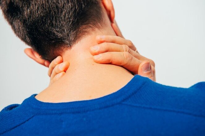
With the osteochondrosis of the spinal column, many are not familiar with popular marches from the TV screen, but by their sad experience.The statistics are hard: up to 80% of the population suffers from this disorder, which also significantly younger.If previous complaints about problems in the spine were mainly among the older generations, now the osteochondosis of children no longer surprises anyone.And the fault of a sedentary lifestyle and the so called "benefits of civilization".

The osteochondrosis of the cervical column is political a progressive disease that occurs through degeneration of the intervertebral discs and dystrophy of the binding system of the spine.Everyone knows the symptoms in the first person, but this knowledge is fragmentary;We will try to structure them, as well as to speak of the principles of diagnosis and treatment of osteochondosis of the cervical column.
The causes of osteochondosis
Medical science cannot answer unequivocally, which is why osteochondrosis occurs.It is reliably known that the sedentary lifestyle that a modern person is subject to negatively influence on the progression of this disease.It is interesting to note that both the hypodynamia and the colossal loads of athletes lead to the proxy of the records.A hereditary factor plays a leading role.The following reasons are distinguished:
- Welled hereditary history;
- obesity;
- Hypodynamia;
- metabolic disorders in the body;
- traumatic damage to the spine;
- long static overloads and works associated with weight lifting (computer work, weight lifting, miners, removals, etc.);
- scoliosis;
- dysfunctional environmental situation;
- Feet dishes and pregnancy;
- Hypothermia and stress, which often cause exacerbations of the disease.
There are several neurological syndromes:
- Shoulder -Shoulder Periarthrite;
- root;
- cardiac;
- Vail artery syndrome.
Shoulder -Shoulder periartrite.It is characterized by neck pain, shoulder, shoulder joint.The main neurogenic contracture of the shoulder joint is formed, which is of a protective nature, as it protects the axillary nerve from lengthening (angle laying).With this position, the muscles surrounding the joint are in tension.The severity of pain syndrome depends on the degree of exacerbation of the osteocondrosis: on a slight limitation of the amplitude of the movements in the articulation to the so called "frozen shoulder", when any movements cause severe pain.The pain intensifies when the shoulder is deflected and pronounced, since these are these movements that improve the tension of the axillary nerve.
Royshift syndrome (cervical radicalitis).More often it occurs with cervical osteochondrosis.At the same time, the spinal nerve spinal column is crushed due to the "subsidia" of the intervertebral discs, as well as due to the growth of osteophytes or the protrusion of the discs in the side direction.Pain syndrome is specific: intense pain, tear, early, which intensifies even when the patient moves his head.The analgical laying is also known in the muscles of the neck, they are strongly tense and painful, the volume of the movements is limited.There is pain in the back of the head, the neck, the front chest, the shoulder, between the shoulder blades.The interruption of sensitivity by the type of "half jacket with short sleeves" is characteristic.
Cardial syndrome.The name of the syndrome is responsible for himself: the clinical image is very similar to the angina pectis.In this case, there is no organic damage to the heart, at the height of pain syndrome, violations of the coronary blood flow by the ACG are not detected and these patients are well tolerated.A typical feature with Angina Pectoris: the pain takes place after taking nitrates and in the case of osteocondrosis does not change and worries for a long time.Unlike the Angina Pectoris, the location of pain is mainly in the heart on the left.With the irritation of the roots of the C8 - T1 segments, rhythmic disorders are possible in the form of tachycardia and extrashole.This is not due to damage to the conductor system of the heart, but with a violation of the sympathetic change of the heart muscle (extracardiac damage).In the differential diagnosis of Angina Pectoris and cardiac syndrome, the main one is the fact that, in addition to the cardial complaints, the patient detects the increase in pain in the shoulder joint and neck associated with lifting or hard movements.
Vail artery syndrome.The vertebral artery takes place in a channel formed by holes in the transversal processes of the vertebrae.This artery is coupled, it is responsible for the blood flow to the brain.Consequently, any narrowing of this channel affects very negatively on the nutrition of the brain tissue.The vertebral artery syndrome develops directly both with the compression of the artery itself and with the irritation of the sympathetic nervous plexus, which is found around it.The pain in this pathology is burning or pulsating in the occipital region with diffusion on whiskey, arches tutorials, crows.It rises on both and on both sides.Patients usually associate aggravation to the condition after sleep in a non -physiological pose, travel in transport, walking.With pronounced symptoms, hearing loss, dizziness, noise in the ears, nausea, vomiting, loss of consciousness and increased blood pressure.These symptoms are not specific and are very similar to complaints in the cerebral stroke.This pathology is characterized by the Sistine chapel syndrome: a fainting that occurs when it reverses the head (severe cerebral ischemia).He was described by the visitors of the Sistine Chapel to the Vatican when they examined the frescoes in his arches.It is also possible to fall without loss of consciousness with acute turns of the head.
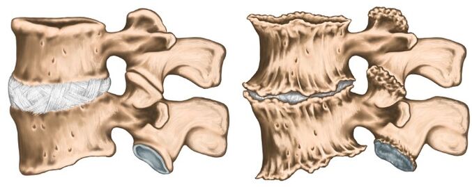
Like any diagnosis in medicine, the diagnosis of osteochondrosis is established on the basis of patients' complaints, anamnesis of the disease, clinical examination and auxiliary research methods.The X -ray of the cervical column is performed in direct and lateral projections, if necessary in special positions (with an open mouth).At the same time, experts are interested in the height of the intervertebral records, in the presence of osteophytes.Of modern research methods, IAMR and CT research are used, which allow you to verify the diagnosis in a more accurate way.In addition to the methods listed in further research, consultations of related specialists (cardiologist, ophthalmologist, neurosurgeon) and the examination of the neurologist is simply vital can be needed.The neurologist is engaged in the treatment of osteocondrosis, therefore after examining the patient, he will prescribe the minimum examination necessary at his discretion.
Treatment of osteocondrosis
Osteochondrosis is a polygiological disease, since a therapy course is not treated.You cannot drink a "magic pill" and everything will pass, it is necessary to radically change your lifestyle, since the trigger is hypodynamia.The most tangible results are easier to achieve in the initial phase of the disease, when the complaints are minimal and there are no compression syndromes and spinal artery.In the acute phase of the disease, when the following groups of drugs are prescribed pronounced pain: pain syndrome is pronounced:
- Therapeutic paravertebral block (to relieve the pain and removal of muscle spasm);
- Fanss;
- ointments containing fans and reflected action;
- muscle relaxants;
- B Vitamins V.
As the inflammatory process mitigates and the relief of pain syndrome, they move on to physiotherapy treatment.Very often the following techniques are used:
- laser therapy;
- electrophoresis;
- acupuncture;
- Operating therapy;
- therapeutic massage;
- Manual therapy.
It is important to understand that osteochondosis proceeds with periods of exacerbation and remission, therefore it is very important to influence the cause and not to treat investigations.